Diabetic Retinopathy
Pathogenesis
Diabetic retinopathy is the result of microvascular retinal changes. Hyperglycemia-induced pericyte death and thickening of the basement membrane lead to incompetence of the vascular walls. These damages change the formation of the blood-retinal barrier and also make the retinal blood vessels become more permeable.
Small blood vessels - such as those in the eye - are especially vulnerable to poor blood sugar (blood glucose) control.
An overaccumulation of glucose and/or fructose damages the tiny blood vessels in the retina. During the initial stage, called nonproliferative diabetic retinopathy (NPDR), most people do not notice any change in their vision.
Some people develop a condition called macular edema. It occurs when the damaged blood vessels leak fluid and lipids onto the macula, the part of the retina that lets us see detail. The fluid makes the macula swell, which blurs vision.
As the disease progresses, severe nonproliferative diabetic retinopathy enters an advanced, or proliferative, stage. The lack of oxygen in the retina causes fragile, new, blood vessels to grow along the retina and in the clear, gel-like vitreous humour that fills the inside of the eye. Without timely treatment, these new blood vessels can bleed, cloud vision, and destroy the retina. Fibrovascular proliferation can also cause tractional retinal detachment. The new blood vessels can also grow into the angle of the anterior chamber of the eye and cause neovascular glaucoma. Nonproliferative diabetic retinopathy shows up as cotton wool spots, or microvascular abnormalities or as superficial retinal hemorrhages. Even so, the advanced proliferative diabetic retinopathy (PDR) can remain asymptomatic for a very long time, and so should be monitored closely with regular checkups. -Source Wikipedia
Diagnosis
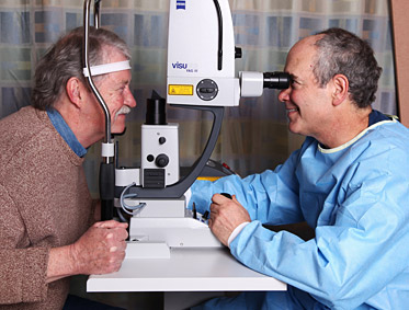
A dilated fundus exam is required to diagnose diabetic retinopathy. This exam should include a visual acuity test, pupil evaluation, eye muscle evaluation, a refraction to determine your best corrected vision, and a evaluation of the outside of the eye and the inside of the eye with a microscope called a slit lamp. In order to see the blood vessels inside the eye properly the pupil needs to be dilated with eye drops.